- Analytical
- Logical
- Verbal
- Language
- Intelligence
- Intuition
- Creativity
- Emotions
- Imagination.
- Rhythm
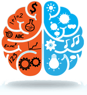
BETTER HEALTH STARTS IN THE BRAIN
WHY BRAIN HEALTH MATTERS
The brain plays an active role in nearly every function of your body. From logic and critical thinking to imagination and talent; from digestion and sleep quality to heart rate and recovery; the brain is there, making sure everything runs smoothly.
So when the brain isn't working properly, it can affect us in many negative ways. When we lose our temper or experience extreme sadness or lack of control, it could very well be that our brain is out of balance. An improperly balanced brain can lead to many neurological problems, such as:
- ADHD & Learning Issues
- Addiction & Abuse
- Anxiety & Stress
- Brain Injury & Concussion
- Chronic Pain & Fatigue
- Depression
- Epilepsy & Seizures
- Insomnia & Sleep Issues
- Memory Loss
- Migraines
- Stroke & More
We Can Help Balance Your Brain!
We offer a
scientifically proven system that can safely fine-tune the brain to eliminate the triggers that often cause the issues above. We can also help over come attention and focus issues, as well as improve
memory retention. Best of all, this process is
drug-free, safe and non-invasive.
What is Neurofeedback?
What if you could eliminate or reduce chronic neurological conditions just by watching a movie or listening to music?
That may sound too good to be true, but this amazing technology works by re-aligning brainwaves while you are engaged in a movie or music. Decades of research have shown that properly aligned brainwaves can positively affect the way our body functions.
How Does It Work?
Neurofeedback does not directly target conditions and symptoms: it corrects irregular brainwaves and modifies timing patterns in the brain. This is achieved over multiple neurofeedback sessions, as the brain is re-trained into normal patterns. The result is an improvement in brain regulation, which can impact a variety of symptoms.
Think of your brain as a musical quartet: When all musicians are in sync, the sound is harmonious. But if one musician is out of tune, the overall sound is affected. Brainwaves operate in much the same way, working together to keep your mind and body in sync and running smoothly. But if any brainwaves are off, it can impact your entire system negatively. Many common conditions like anxiety, depression and others can occur when brainwaves are running too fast or too slow. Neurofeedback teaches the brain to regulate its brainwaves properly, which can result in better overall health.
Neurofeedback Is Not New
It’s been around since the 1960’s. There are decades of research and case studies that document its effectiveness in improving brain health. Advances in computer technology have made it possible for doctors to easily administer neurofeedback in their clinic.
How Does It Work?
Neurofeedback has 3 main goals:
- Identify irregular brainwaves (Alpha, Beta, Delta and Theta)
- Guide those brainwaves back into regular patterns
- Teach the brain to maintain regular patterns permanently
Neurofeedback works primarily by monitoring brainwaves on the surface of your head. To start, small electrodes are placed on your scalp. These electrodes have a paste on them which makes it easier to pick up brainwave patterns. For the next 30 minutes, you get to watch a movie of your choice, listen to your music or listen to an audio book. That is all that is required of you.
Is is non-invasive, uses no drugs and does not involve any radiation.
During a normal session the computer is monitoring your brainwaves, looking for any that are out of the normal range. When it finds one, the system triggers a response that changes the movie or music. This change is not annoying, but it is subtle enough to get your attention and make you focus more. Refocusing corrects the irregular brainwaves, which then move into the normal range. At that time the movie or music will resume normally. This process is called Operant Conditioning. Over the course of multiple sessions, the brain eventually learns to make healthy patterns on it's own. At that time, no further neurofeedback sessions are needed.
What is Brain Mapping?
Brain Map: A Customized, Accurate Analysis Of Your Brain?
A Brain Map is a non-invasive tool we use to identify the problem areas of the brain. There is no more accurate tool available today for identifying irregular brainwaves. It also generates a set of protocols that can correct your specific brainwave irregularities using neurofeedback and other modalities. The Brain Map process is
painless, safe, accurate and non-invasive. There really is no better tool for analyzing brainwaves and collecting customized data for each individual.
How Does A Brain Map Work?
A brain map involves scanning the brainwaves on the surface of the scalp using a nylon cap. This method is known as an Quantitative Electroencephalogram (QEEG) and provides the
most accurate recording of your normal brain function. The system then compares your brainwave activity to a database of established standards of normal brain function to determine if problems are present. It does not identify specific conditions: It shows a map of problem areas in the brain that we can use to expertly determine likely neurological conditions.
Why Brainwaves Are Important
Brainwaves are extremely important to how we function. There are 4 main brainwaves, and each of them regulates a different part of our body. From sleep to emotions to critical thinking, we would not be who we are without our brainwaves. Let's learn about each one.
Beta Waves
These brainwaves are commonly observed while we are awake. They are involved in conscious thought, logical thinking, writing, reading and stimulation. Having the right amount of beta waves allows us to focus and complete school or work-based tasks easily. Having too much beta may lead to us experiencing excessive stress and/or anxiety.
Alpha Waves
This frequency range bridges the gap between our conscious thinking and subconscious mind. It helps us calm down when necessary and promotes feelings of deep relaxation. If we become stressed, a phenomenon called “alpha blocking” may occur which involves the beta waves “blocking” the production of alpha waves.
Theta Waves
This particular frequency range is involved in daydreaming and sleep. Theta waves are connected to us experiencing and feeling deep and raw emotions. Too much theta activity may cause depression and make people “highly suggestible” because they are in a deeply relaxed, semi-hypnotic state. Theta can improve intuition, creativity, and makes us feel more natural. It is also involved in restorative sleep.
Delta Waves
These are the slowest recorded brain waves in human beings. They are associated with the deepest levels of relaxation and restorative, healing sleep. Adequate production of delta waves helps us feel completely rejuvenated after we wake up from a good night’s sleep.
The Brain Map Report
What makes our system so unique and successful is the ability to analyze each brain and generate a customized report that shows the problem areas of a persons brain. This comprehensive report of findings is unique to each individual and very detailed. Not only does it show problem areas, but also how to improve them with neurofeedback. Think of it as a customized care plan for your brain.
Frequently Asked Questions
What Happens During a Neurofeedback Session?
Neurofeedback sessions involve relaxing for 30 minutes while you watch a movie or listen to music of your choice. electrodes are attached to your scalp that monitor your brainwaves during the session. When irregular patterns are detected, a response is triggered from the software that pauses or dims the video or music. Your brain senses the change and subconsciously modifies itself back into a normal pattern. With repetition of this process, eventually your brain learns to stay within healthy ranges on its own without neurofeedback.
How Long do Neurofeedback Sessions take?
Each session is 30 minutes.
How many neurofeedback sessions are needed?
The number of sessions needed will depend on the individual. Much like going to the gym, every person requires a different length of time to improve. 20 – 40 sessions is normal for many conditions to improve.
How soon will I see results from Neurofeedback?
Again, results will vary from person to person. Some feel different within a couple of sessions, while tougher conditions will take many sessions to see any noticeable results. It’s important to not get impatient and listen to the practitioner. They should be able to show you the graph results of each sessions, which will provide a visual reference of improvement.
How Long Will the Effects Of Neurofeedback Last?
Long term follow ups have been done on many patients over the years. Dr. Joel Lubar at the University of Tennessee has followed ADD clients who’ve sustained their improvements from neurofeedback for 10-20 years. Published research on epilepsy 12 months after brain training shows the effects on epilepsy usually holds. Owners of the Clear Mind System have commonly reported no relapses from patients after 10 years.
How much research is there on Neurofeedback?
Neurofeedback has been around for decades. To date there are thousands of studies, with more being published every day. This site has a comprehensive list of studies on neurofeedback for many conditions. You can view them here.
Testimonials
Lorna and Fibromyalgia
Laurie & Anxiety
RESEARCH ARTICLES

People with ADD can have a variety of symptoms. They can be easily distracted, impulsive, and inattentive However, ADD is not laziness or a psychological problem – it's a brain problem. Doctors know ADD is not laziness; that's why they prescribe medications. Unlike medication, neurofeedback trains the brain, resulting in significant improvement in ADHD/ADD symptoms, With neurofeedback, people can increase self-control and attention. According to health professionals who use neurofeedback in their practices, many clients with ADD/ADHD learn to increase focus, reduce impulsivity, and manage their behavior when they train with neurofeedback on a consistent basis.
Evidence-Based Information on the Clinical Use of Neurofeedback for ADHD [pdf]
Tais S. Moriyama, Guilherme Polanczyk, and Luis A. Rohde www.ncbi.nlm.nih.gov/pmc/articles/PMC3441929/
Neurofeedback (NF) is a training to enhance self-regulatory capacity over brain activity patterns and consequently over brain mental states. Recent findings suggest that NF is a promising alternative for the treatment of attention-deficit/hyperactivity disorder (ADHD). We comprehensively reviewed literature searching for studies on the effectiveness and specificity of NF for the treatment of ADHD. In addition, clinically informative evidence-based data are discussed. We found 3 systematic review on the use of NF for ADHD and 6 randomized controlled trials that have not been included in these reviews. Most nonrandomized controlled trials found positive results with medium-to-large effect sizes, but the evidence for effectiveness are less robust when only randomized controlled studies are considered. The direct comparison of NF and sham-NF in 3 published studies have found no group differences, nevertheless methodological caveats, such as the quality of the training protocol used, sample size, and sample selection may have contributed to the negative results. Further data on specificity comes from electrophysiological studies reporting that NF effectively changes brain activity patterns. No safety issues have emerged from clinical trials and NF seems to be well tolerated and accepted. Follow-up studies support long-term effects of NF. Currently there is no available data to guide clinicians on the predictors of response to NF and on optimal treatment protocol. In conclusion, NF is a valid option for the treatment for ADHD, but further evidence is required to guide its use.

Neurofeedback training has been used with several thousand autistic spectrum children over the last 15 years, by hundreds of clinicians. There have been several research studies published to support these efforts. What's the first thing parents consistently report as their children start training? They usuall notice their child is more calm, manages emotions better, and doesn't get overwhelmed as easily. There are many other changes, as noted below, but these are typically the first.
QEEG Characteristics and Spectrum Weighted Frequency for Children Diagnosed as Autistic Spectrum Disorder [pdf]
Nada Pop-Jordanova, Tatjana Zorcec, Aneta Demerdzieva, Zoran Gucev Pop-Jordanova et al. Nonlinear Biomedical Physics 2010
Background:Autistic spectrum disorders are a group of neurological and developmental disorders associated with social, communication, sensory, behavioral and cognitive impairments, as well as restricted, repetitive patterns of behavior, activities, or interests. The aim of this study was a) to analyze QEEG findings of autistic patients and to compare the results with data base; and b) to introduce the calculation of spectrum weighted frequency (brain rate) as an indicator of general mental arousal in these patients. Results: Results for Q-EEG shows generally increased delta-theta activity in frontal region of the brain. Changes in QEEG pattern appeared to be in a non-linear correlation with maturational processes. Brain rate measured in CZ shows slow brain activity (5. 86) which is significantly lower than normal and corresponds to low general mental arousal. Recent research has shown that autistic disorders have as their basis disturbances of neural connectivity. Neurofeedback seems capable of remediating such disturbances when these data are considered as part of treatment planning. Conclusions: Prognosis of this pervasive disorder depends on the intellectual abilities: the better intellectual functioning, the possibilities for life adaptation are higher QEEG shows generally increased delta-theta activity in frontal region of the brain which is related to poor cognitive abilities. Brain rate measured in CZ shows slow brain activity related to under arousal. Pharmacotherapy combined with behavior therapy, social support and especially neurofeedback technique promise slight improvements.

Brain training via neurofeedback teaches the brain to maintain a consistent state. Learning self-regulation allows a person to achieve mood stabilization. After beginning neurofeedback, clients commonly comment that they can once again "trust their brain." What does this mean? Bipolar clients undergoing neurofeedback training report less susceptibility to mood swings, increased ability to focus, and reduced anger. Their ability to function increases as they find themselves less reactive and increasingly able to respond and act appropriately.
The Bipolar Child
by Demitri and Janice Papolos Book review by Siegfried Othmer, Ph.D
A new diagnostic category is emerging: Childhood bipolar disorder. It was traditionally thought that as few as one in 200 cases of bipolar disorder had an onset which could be traced to childhood. Biederman's recent research shows that perhaps on the other of 20% of children identified as ADHD could be on the way to developing full-blown bipolar disorder. To make this identification, however, the markers of childhood bipolar disorder are destructive rage and irritation rather than the euphoria and elation that characterizes the adult form. The proof that the childhood form of the disorder metamorphoses into the adult form eventually must still be outstanding. The model is still too new. The Bipolar Disorder model is the latest attempt to give diagnostic order and specificity to the most extreme end of the disruptive behavior spectrum. It is of course not the first. Years ago, George Murray of Harvard suggested that temporal lobe epilepsy was under-recognized by mental health professionals by a factor of 25. Clearly he was not referring to overt seizures here, which tend to attract clinical attention, but rather to the subclinical seizure activity that can manifest in erratic behavior, severe mood swings, rages and explosive behavior - but goes unrecognized as such. Partly based on Murray's model, we have emphasized as well the continuity between overt seizures and extreme behavioral disregulation. Both are effectively treated with anti-convulsants, and both respond to the same Neurofeedback protocols. The developments in Neurofeedback therapy neatly parallel developments in psychopharmacology. But seizures have remained in the domain of neurology, and other mental health professionals have been reluctant to build on that model.

With a traumatic brain injury (TBI), the brain itself needs to be targeted. With neurofeedback, the brain is exercised. The specific areas of the brain affected by the TBI are targeted during neurofeedback therapy. Often in the case of TBI, a neurofeedback practitioner will utilize a qEEG brain map to determine which areas should be targeted. A variety of symptoms can be improved through neurofeedback training, such as speech, movement, regulating moods, controlling behavior, and reducing headaches. Neurofeedback works because the brain regulates each of those issues. For people recovering from TBI, neurofeedback training can be particularly helpful in improving speech. During neurofeedback training, the specific areas of the brain related to speech can be targeted. In this way, the areas associated with speech can be strengthened and improved. In fact, some neuropsychologists believe that neurofeedback is actually rehabilitating the damaged speech areas of the brain rather than just dealing with compensation.
Evaluation of Differentiated Neurotherapy Programs for a Patient After Severe TBI and Long Term Coma Using Event-related Potentials
Maria Pachalska1, Małgorzata Łukowicz, Juri D. Kropotov, Izabela Herman-Sucharska, Jan Talar The Medical Science Monitor, 2011
BackgroundThis article examines the effectiveness of differentiated rehabilitation programs for a patient with frontal syndrome after severe TBI and long-term coma. We hypothesized that there would be a small response to relative beta training, and a good response to rTMS, applied to regulate the dynamics of brain function. Case Report M. L-S, age 26, suffered from anosognosia, executive dysfunction, and behavioral changes, after a skiing accident and prolonged coma, rendering him unable to function independently in many situations of everyday life. Only slight progress was made after traditional rehabilitation. The patient took part in 20 sessions of relative beta training (program A) and later in 20 sessions of rTMS (program B); both programs were combined with behavioral training. We used standardized neuropsychological testing, as well as ERPs before the experiment, after the completion of program A, and again after the completion of program B. As hypothesized, patient M.L-S showed small improvements in executive dysfunction and behavioral disorders after the conclusion of program A, and major improvement after program B. Similarly, in physiological changes the patient showed small improvement after relative beta training and a significant improvement of the P300 NOGO component after the rTMS program. Conclusions The rTMS program produced larger physiological and behavioral changes than did relative beta training. A combination of different neurotherapeutical approaches (such as neurofeedback, rTMS, tDCS) can be suggested for similar severe cases of TBI. ERPs can be used to assess functional brain changes induced by neurotherapeutical programs.

Pain is one of several sensory systems that keep us apprised of the status of our bodies. As we hurry through our daily lives, we usually view pain at the very least as an inconvenience, if not a major disruption. It's fortunate that we have our pain sensors-they provide a valuable warning to us that we need to stop and take care of ourselves. For chronic pain, neurofeedback can help reduce pain or perhaps how the brain manages pain, even in severe cases.
New Hope for Sufferers of Chronic Pain [pdf]
by Siegfried Othmer, Ph.D.
Pain is one of several sensory systems that keep us apprised of the status of our bodies. As we hurry through our daily lives, we usually view pain at the very least as an inconvenience, if not a major disruption. It's fortunate that we have our pain sensors - they provide a valuable warning to us that we need to stop and take care of ourselves. Pain has considerable survival value, but when an injury has healed and the pain continues unabated, or when pain seems to have no obvious connection to any injury, it no longer serves a useful purpose. Pain of this type is referred to as chronic pain, and once you have fallen under its sway, it may be very difficult to escape. The Challenge of Pain Management The management of chronic pain has always been a medical challenge. Treatment often involves increasing doses of a variety of medications in an effort to gain a measure of relief. In some instances, the pain is significantly reduced with the use of medication, but when the drugs are removed the pain returns, and so the meds become a more or less permanent fixture of life, often resulting in drug dependence or even addiction. In other cases even heavy use of medication provides the sufferer little or no relief; the brain simply adjusts to the presence of the medications and demands more, while the pain continues.

Feeling down or depressed from time to time happens to most people. Usually such feelings pass, and a person can improve his or her mood naturally. However, some people cannot break out of a depressed state over an extended period of time. In those cases, a person is considered to have clinical depression. However, there is much research that shows that depression is neurological, not psychological. Certain brain patterns are frequently linked to depression. Therefore, training the brain through neurofeedback has a powerful ability to treat depression. With neurofeedback training, the brain practices a healthy pattern of mood regulation. Sometimes people with depression notice improvement after only a few sessions. However, for the brain to fully learn, more training is required. In time, the brain learns to regulate mood on its own.
Real-Time Self-Regulation of Emotion Networks in Patients with Depression [pdf]
David E. J. Linden, Isabelle Habes, Stephen J. Johnston, Stefanie Linden, Ranjit Tatineni, Leena Subramanian, Bettina Sorger, David Healy1, Rainer Goebe
AbstractMany patients show no or incomplete responses to current pharmacological or psychological therapies for depression. Here we explored the feasibility of a new brain self-regulation technique that integrates psychological and neurobiological approaches through neurofeedback with functional magnetic resonance imaging (fMRI). In a proof-of-concept study, eight patients with depression learned to upregulate brain areas involved in the generation of positive emotions (such as the ventrolateral prefrontal cortex (VLPFC) and insula) during four neurofeedback sessions. Their clinical symptoms, as assessed with the 17-item Hamilton Rating Scale for Depression (HDRS), improved significantly. A control group that underwent a training procedure with the same cognitive strategies but without neurofeedback did not improve clinically. Randomized blinded clinical trials are now needed to exclude possible placebo effects and to determine whether fMRI-based neurofeedback might become a useful adjunct to current therapies for depression.

A seizure disorder can be explained as a brain that has lost stability. People with seizures can regulate and stabilize their brains through neurofeedback training. Eighteen well-run research studies show how effective neurofeedback training can be in the reduction of seizures. Interestingly, this research began with studies performed on cats. In an experiment to determine neurofeedback's effectiveness to combat seizures, it was found that cats with neurofeedback training, when exposed to a chemical, experienced far fewer seizures than those without the training.
A model of feedback control for the charge-balanced suppression of epileptic seizures [link]
Beth A. Lopour and Andrew J. Szericorresponding Journal of Computational Neuroscience, (2010)
AbstractHere we present several refinements to a model of feedback control for the suppression of epileptic seizures. We utilize a stochastic partial differential equation (SPDE) model of the human cortex. First, we verify the strong convergence of numerical solutions to this model, paying special attention to the sharp spatial changes that occur at electrode edges. This allows us to choose appropriate step sizes for our simulations; because the spatial step size must be small relative to the size of an electrode in order to resolve its electrical behavior, we are able to include a more detailed electrode profile in the simulation. Then, based on evidence that the mean soma potential is not the variable most closely related to the measurement of a cortical surface electrode, we develop a new model for this. The model is based on the currents flowing in the cortex and is used for a simulation of feedback control. The simulation utilizes a new control algorithm incorporating the total integral of the applied electrical potential. Not only does this succeed in suppressing the seizure-like oscillations, but it guarantees that the applied signal will be charge-balanced and therefore unlikely to cause cortical damage.

Fibromyalgia is "A common syndrome of chronic widespread soft-tissue pain accompanied by weakness, fatigue, and sleep disturbances; the cause is unknown." The word fibromyalgia comes from the Greek myos meaning "muscle", Greek algos meaning "pain", and New Latin fibro meaning "fibrous tissue". Fibromyalgia is a common and chronic disorder. When a health illness or condition is chronic it means it is long-lasting. Even though fibromyalgia is frequently referred to as an arthritis-related condition, it does not cause joint damage or inflammation, as arthritis does. Neither does fibromyalgia cause damage to muscle and other tissues. However, it is similar to arthritis because it causes severe.
Efficacy of EMG- and EEG-Biofeedback in Fibromyalgia Syndrome: A Meta-Analysis and a Systematic Review of Randomized Controlled Trials [link]
Julia Anna Glombiewski, Kathrin Bernardy and Winfried Häuser www.ncbi.nlm.nih.gov/pmc/articles/PMC3776543/
AbstractBiofeedback (BFB) is an established intervention in the rehabilitation of headache and other pain disorders. Little is known about this treatment option for fibromyalgia syndrome (FMS). The aim of the present review is to integrate and critically evaluate the evidence regarding the efficacy of biofeedback for FMS. Methods. We conducted a literature search using Pubmed, clinicaltrials.gov (National Institute of Health), Cochrane Central Register of Controlled Trials, PsycINFO, SCOPUS, and manual searches. The effect size estimates were calculated using a random-effects model. Results. The literature search produced 123 unique citations. One hundred sixteen records were excluded. The meta-analysis included seven studies (321 patients) on EEG-Biofeedback and EMG-Biofeedback. In comparison to control groups, biofeedback (BFB) significantly reduced pain intensity with a large effect size (g = 0.79; 95% CI: 0.22–1.36). Subgroup analyses revealed that only EMG-BFB and not EEG-BFB significantly reduced pain intensity in comparison to control groups (g = 0.86; 95% CI: 0.11–1.62). BFB did not reduce sleep problems, depression, fatigue, or health-related quality of life in comparison to a control group. Discussion. The interpretation of the results is limited because of a lack of studies on the long-term effects of EMG-BFB in FMS. Further research should focus on the long-term efficacy of BFB in fibromyalgia and on the identification of predictors of treatment response.

Many of the methods used and promoted to help people with learning disabilities are intended to help a person compensate for, or work around, their learning difficulties. Neurofeedback actually improves learning skills by training the areas of the brain relevant to learning or executing skills such as math, reading, and auditory and visual processing. Research studies show that several areas of the brain coordinate in the learning process. These separate parts of the brain communicate with each other at extremely fast speeds. If the timing of the communication is even slightly off, there can be impairment in the ability to learn. New research shows that this "connectivity training" seems to consistently improve learning difficulties. Neurofeedback training can improve the coordination and communication between different areas of the brain. Improved timing in the brain has a significant impact on one's ability to learn. Neurofeedback directly targets the coordination and communication between areas of the brain to improve timing, and therefore learning. pain and tiredness, and can undermine the patient's ability to go about his daily activities. Fibromyalgia is seen as a rheumatic condition. A rheumatic condition is one that causes joint and soft tissue pain.
Research Review: Emanuel Miller Memorial Lecture 2012 – Neuroscientific studies of intervention for language impairment in children: interpretive and methodological problems [link]
D V M Bishop www.ncbi.nlm.nih.gov/pmc/articles/PMC3593170/
BackgroundOur ability to look at structure and function of a living brain has increased exponentially since the early 1970s. Many studies of developmental disorders now routinely include a brain imaging or electrophysiological component. Amid current enthusiasm for applications of neuroscience to educational interventions, we need to pause to consider what neuroimaging data can tell us. Images of brain activity are seductive, and have been used to give credibility to commercial interventions, yet we have only a limited idea of what the brain bases of language disorders are, let alone how to alter them. Scope and findings A review of six studies of neuroimaging correlates of language intervention found recurring methodological problems: lack of an adequate control group, inadequate power, incomplete reporting of data, no correction for multiple comparisons, data dredging and failure to analyse treatment effects appropriately. In addition, there is a tendency to regard neuroimaging data as more meaningful than behavioural data, even though it is behaviour that interventions aim to alter. Conclusion In our current state of knowledge, it would be better to spend research funds doing well-designed trials of behavioural treatment to establish which methods are effective, rather than rushing headlong into functional imaging studies of unproven treatments.

Although neurofeedback training can stop a migraine while it is occurring, stopping individual migraines is not the main goal. Training with neurofeedback can be very effective in reducing the intensity and frequency of migraines over the long term providing real relief for people suffering from migraines. Deborah Stokes, Ph.D, a neurofeedback clinician in Alexandria, VA. recently published a study that showed significant improvement in migraines using neurofeedback. The study was co-authored with Martha S. Lappin and entitled "Neurofeedback and biofeedback with 37 migraineurs: a clinical outcome study". The study found that, with neurofeedback, 70% of migraine sufferers have a significant reduction in the frequency of their migraines.
Neurofeedback and biofeedback with 37 migraineurs: a clinical outcome study [pdf]
Deborah A Stokes, Martha S Lappin Behavioral and Brain Functions 2010, 6:9
Background:Traditional peripheral biofeedback has grade A evidence for effectively treating migraines. Two newer forms of neurobiofeedback, EEG biofeedback and hemoencephalography biofeedback were combined with thermal handwarming biofeedback to treat 37 migraineurs in a clinical outpatient setting. Methods: 37 migraine patients underwent an average of 40 neurofeedback sessions combined with thermal biofeedback in an outpatient biofeedback clinic. All patients were on at least one type of medication for migraine; preventive, abortive or rescue. Patients kept daily headache diaries a minimum of two weeks prior to treatment and throughout treatment showing symptom frequency, severity, duration and medications used. Treatments were conducted an average of three times weekly over an average span of 6 months. Headache diaries were examined after treatment and a formal interview was conducted. After an average of 14.5 months following treatment, a formal interview was conducted in order to ascertain duration of treatment effects.

With Obsessive Compulsive Disorder (OCD), a person can't stop repeating specific behaviors or stop his or her brain from repeating particular thoughts. A substantial body of research shows that problems with OCD are related to the functioning of areas in the front of the brain. If that part of the brain is working too slowly or quickly, a person is unable to stop repeating certain thoughts or behaviors. Many therapists and other health professionals using neurofeedback to treat OCD note marked reductions in OCD symptoms in their clients after neurofeedback training. People with OCD relate that, after neurofeedback training, they do not really need to make an effort to stop unwanted repetitive thoughts and behaviors. They say that they their minds are much quieter. With neurofeedback training, the brain learns to respond to situations in a more conventional and healthy manner.
Obsessive Compulsive Disorder and the Efficacy of qEEG-Guided Neurofeedback Treatment: A Case Series [pdf]
Tanju Siirmeli and Ayben Exrteme Clinical EEG & Neuroscience, Volume 42 No 3
ABSTRACTWhile neurofeedback has been extensively studied in the treatment of many disorders, there have been only three published reports, by D.C. Hammond, on its clinical effects in the treatment of obsessive compulsive disorder (OCD). In this paper the efficacy of QEEG-guided neurofeedback for subjects with OCD was studied as a case series. The goal was to examine the clinical course of the OCD symptoms and assess the efficacy of QEEG guided neurofeedback training on clinical outcome measures. Thirty-six drug resistant subjects with OCD were assigned to 9-84sessions of QEEG-guided neurofeedback treatment. Daily sessions lasted 60minutes where 2 sessions with half-hour applications with a 30 minute rest given between sessions were conducted per day. Thirty-three outof36 subjects who received neurofeedback training showed clinical improvement according to the Yale-Brown obsessive-compulsive scale (Y-BOCS). The Minnesota multiphasic inventory(MMPI) was ad-ministered before and after treatment to 17 of the subjects. The MMPI results showed significant improvements not only in OCD measures, but all of the MMP1 scores showed a general decrease. Finally ,according to the physicians' evaluation of the subjects using the clinical global impression scale (CGI), 33 of the 36 subjects were rated as improved. Thirty-six of the subjects were followed for an average of 26months after completing the study. According to follow-up interviews conducted with them and/or their family members 19of the subjects maintained the improvements in their OCD symptoms. This study provides good evidence for the efficacy of neurofeedback treatment in OCD. The results of this study encourage further controlled research in this area.

Post Traumatic Stress Disorder (PTSD) is a serious type of anxiety caused by an extremely stressful event or series of events. People who suffer from PTSD are looking for a method to treat their symptoms, and unfortunately, many people experience only limited benefit after trying various therapies and medication. Neurofeedback trains the brain to produce a calm state as well as regulate stress response. In addition, the specific areas of the brain affected by PTSD can be targeted. Frequently, the first sign of improvement is that a client sleeps better. Then other symptoms begin to improve. After sufficient training, someone with PTSD can maintain a calm state on his or her own. When a person has reached this stable state, neurofeedback treatments can be decreased until no further trainings are necessary.
The long-term costs of traumatic stress: intertwined physical and psychological consequences [pdf]
Alexander C. McFarlane
ABSTRACTThe gradual emergence of symptoms following exposure to traumatic events has presented a major conceptual challenge to psychiatry. The mechanism that causes the progressive escalation of symptoms with the passage of time leading to delayed onset post-traumatic stress disorder (PTSD) involves the process of sensitization and kindling. The development of traumatic memories at the time of stress exposure represents a major vulnerability through repeated environmental triggering of the increasing dysregulation of an individual's neurobiology. An increasing body of evidence demonstrates how the increased allostatic load associated with PTSD is associated with a significant body of physical morbidity in the form of chronic musculoskeletal pain, hypertension, hyperlipidaemia, obesity and cardiovascular disease. This increasing body of literature suggests that the effects of traumatic stress need to be considered as a major environmental challenge that places individual's physical and psychological health equally at risk. This broader perspective has important implications for developing treatments that address the underlying dysregulation of cortical arousal and neurohormonal abnormalities following exposure to traumatic stress.

Schizophrenia is a mental disorder that generally appears in late adolescence or early adulthood – however, it can emerge at any time in life. It is one of many brain diseases that may include delusions, loss of personality (flat affect), confusion, agitation, social withdrawal, psychosis, and bizarre behavior. It may be hard to make sense of what a person with schizophrenia is talking about. In some cases, the individual may spend hours completely still, without talking. On other occasions he or she may seem fine, until they start explaining what they are truly thinking. according to the National Institute of Mental Health (NIMN), treatment can help relieve many of the symptoms of schizophrenia.
Taking Back the Brain: Could Neurofeedback Training Be Effective for Relieving Distressing Auditory Verbal Hallucinations in Patients With Schizophrenia? [link]
Simon McCarthy-Jones www.ncbi.nlm.nih.gov/pmc/articles/PMC3406539/
ABSTRACTProgress in identifying the neural correlates of auditory verbal hallucinations (AVHs) experienced by patients with schizophrenia has not fulfilled its promise to lead to new methods of treatments. Given the existence of a large number of such patients who have AVHs that are refractory to traditional treatments, there is the urgent need for the development of new effective interventions. This article proposes that the technique of neurofeedback may be an appropriate method to allow the translation of pure research findings from AVH-research into a clinical intervention. Neurofeedback is a method through which individuals can self-regulate their neural activity in specific neural regions/frequencies, following operant conditioning of their intentional manipulation of visually presented real-time feedback of their neural activity. Four empirically testable hypotheses are proposed as to how neurofeedback may be employed to therapeutic effect in patients with AVHs.

At least 40 million Americans each year suffer from chronic, long-term, sleep disorders. An additional 20 million experience occasional sleep problems. Neurofeedback is a powerful tool for helping people fall asleep and stay asleep. Over 3,000 licensed health professionals such as psychologists, therapists, and doctors now use this new technology daily with patients. As a group, they report significant and consistent improvements for client sleep problems. Many brain training options can help as well as making lifestyle changes and changes in sleep "hygiene". A skilled neurofeedback clinician can review many different options with clients to help them assess what's most appropriate for their problem, including several brain regulating technologies such as Alpha-Stim and Brain Music.
Neurofeedback in ADHD and insomnia: Vigilance stabilization through sleep spindles and circadian networks. [link]
Arns M, Kenemans JL. www.ncbi.nlm.nih.gov/pubmed/23099283
AbstractIn this review article an overview of the history and current status of neurofeedback for the treatment of ADHD and insomnia is provided. Recent insights suggest a central role of circadian phase delay, resulting in sleep onset insomnia (SOI) in a sub-group of ADHD patients. Chronobiological treatments, such as melatonin and early morning bright light, affect the suprachiasmatic nucleus. This nucleus has been shown to project to the noradrenergic locus coeruleus (LC) thereby explaining the vigilance stabilizing effects of such treatments in ADHD. It is hypothesized that both Sensori-Motor Rhythm (SMR) and Slow-Cortical Potential (SCP) neurofeedback impact on the sleep spindle circuitry resulting in increased sleep spindle density, normalization of SOI and thereby affect the noradrenergic LC, resulting in vigilance stabilization. After SOI is normalized, improvements on ADHD symptoms will occur with a delayed onset of effect. Therefore, clinical trials investigating new treatments in ADHD should include assessments at follow-up as their primary endpoint rather than assessments at outtake. Furthermore, an implication requiring further study is that neurofeedback could be stopped when SOI is normalized, which might result in fewer sessions.

A stroke is a condition in which the brain cells suddenly die because of a lack of oxygen. A stroke can be caused by an obstruction in the blood flow, or the rupture of an artery that feeds the brain. The patient may suddenly lose the ability to speak, there may be memory problems, or one side of the body can become paralyzed.
Parietofrontal integrity determines neural modulation associated with grasping imagery after stroke [pdf]
Ethan R. Buch,Amirali Modir Shanechi, Alissa D. Fourkas, Cornelia Weber, Niels Birbaumer, and Leonardo G. Cohen Brain: A Journal Of Neurology 2012
ABSTRACTChronic stroke patients with heterogeneous lesions, but no direct damage to the primary sensorimotor cortex, are capable of longitudinally acquiring the ability to modulate sensorimotor rhythms using grasping imagery of the affected hand. Volitional modulation of neural activity can be used to drive grasping functions of the paralyzed hand through a brain–computer interface. The neural substrates underlying this skill are not known. Here, we investigated the impact of individual patient's lesion pathology on functional and structural network integrity related to this volitional skill. Magnetoencephalography data acquired throughout training was used to derive functional networks. Structural network models and local estimates of extralesional white matter microstructure were constructed using T1-weighted and diffusion-weighted magnetic resonance imaging data. We employed a graph theoretical approach to characterize emergent properties of distributed interactions between nodal brain regions of these networks. We report that inter-individual variability in patients' lesions led to differential impairment of functional and structural network characteristics related to successful post-training sensorimotor rhythm modulation skill. Patients displaying greater magnetoencephalography global cost-efficiency, a measure of information integration within the distributed functional network, achieved greater levels of skill. Analysis of lesion damage to structural network connectivity revealed that the impact on nodal betweenness centrality of the ipsilesional primary motor cortex, a measure that characterizes the importance of a brain region for integrating visuomotor information between frontal and parietal cortical regions and related thalamic nuclei, correlated with skill. Edge betweenness centrality, an analogous measure, which assesses the role of specific white matter fibre pathways in network integration, showed a similar relationship between skill and a portion of the ipsilesional superior longitudinal fascicle connecting premotor and posterior parietal visuomotor regions known to be crucially involved in normal grasping behaviour. Finally, estimated white matter microstructure integrity in regions of the contralesional superior longitudinal fascicle adjacent to primary sensorimotor and posterior parietal cortex, as well as grey matter volume co-localized to these specific regions, positively correlated with sensorimotor rhythm modulation leading to successful brain–computer interface control. Thus, volitional modulation of ipsilesional neural activity leading to control of paralyzed hand grasping function through a brain–computer interface after longitudinal training relies on structural and functional connectivity in both ipsilesional and contralesional parietofrontal pathways involved in visuomotor information processing. Extant integrity of this structural network may serve as a future predictor of response to longitudinal therapeutic interventions geared towards training sensorimotor rhythms in the lesioned brain, secondarily improving grasping function through brain–computer interface applications.